Speakers
Prof. Dr. med. Pascal Berberat
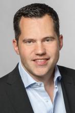
berberat@tum.de
http://www.mec.med.tum.de/de/prof-dr-med-pascal-berberat
The TUM Medical Education Center organizes the education at the faculty of medicine. We are active in two research areas. In the field of instruction and teacher research our focus lies on a better understanding of teaching and learning processes in order to optimize evidence based didactic methods and to systematically analyze the attitude, motivation and behavior of medical doctors as lecturers and tutors. In our second research area we concentrate on medical educational biographies with the main focus on so called ‘non-rational’ dimensions, such as handling existential borderline experiences (suffering, dying and death) und tolerating uncertainty.
Prof. Dr. Arthur Konnerth
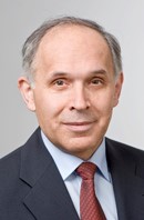
arthur.konnerth@tum.de
http://www.ifn.me.tum.de/new/
Cellular mechanisms of cortical function in vivo: We use two-photon calcium imaging in different areas of the mouse cortex (visual, auditory, sensorymotor) combined with targeted patch-clamp recordings to study electrical signaling and plasticity of specific types of neurons in behaviorally-defined conditions. Cerebellar function and plasticity: We are interested in synaptic mechanisms, including the roles of mGlu receptors, TRPC channels, calcium signaling as well as in cerebellar sensory integration. Dendritic signaling in vivo: Our major aim is the visualization and mapping of sensory-evoked signals on the level of individual synaptic inputs in defined neurons of the mouse cortex. In vivo neurophysiology of Alzheimer’s disease. We focus on the impairments in synaptic signaling of cortical and hippocampal neurons in mouse models of Alzheimer’s disease. Development of imaging technology: We develop and implement two-photon imaging devices with a high spatial and temporal resolution for the functional analysis of networks, cells and subcelullar compartments in vitro and in vivo.
Prof. Dr. Thomas Misgeld
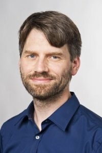
thomas.misgeld@tum.de
https://web.med.tum.de/neuroscience/misgeld-lab/
The Misgeld lab studies axon changes in the healthy and in the sick nervous system of living animals. Axons are the long neuronal processes that form synapses and thus interconnect different parts of the nervous system. Obviously, to properly establish wiring in the brain, myriads of axons have to find their targets, or otherwise, axons that connect incorrectly need to be removed. We are interested in the latter process – not only because such axon dismantling contributes fundamentally to brain development and to the adaptation of our neural circuits to the environment, but also because axons are highly susceptible to pathology. Many common neurological diseases are characterized by early loss of axonal connections – including motor neuron disease, spinal cord injury and multiple sclerosis, all of which we study. By better understanding axon dismantling in development and disease we hope to gain insight into what causes axons to disintegrate in disease.
Faculty
Prof. Dr. Helmuth Adelsberger
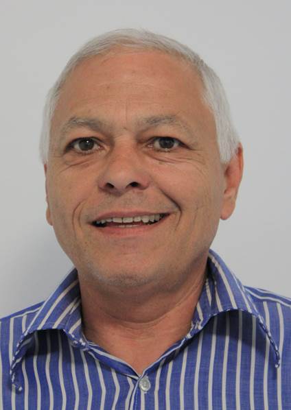
h.adelsberger@tum.de
http://www.ifn.me.tum.de/new/
We develop and use systems based on CCD-cameras and optic fibers for the fluorometric detection of population calcium signals. Such signals are generated by synchronous activity of large numbers of neurons organized in networks. Our recording systems are applied for studying basic brain function and the processing of sensory information as well as for the detection of impairments in mouse models of Alzheimer´s disease. Another focus is the analysis of behavioral impairments in different transgenic mouse models such as models of Alzheimer´s disease or mice with cell-type specific knock-outs in cerebellar Purkinje cells. Furthermore we analyze the effects of drug treatments in our disease mouse models.
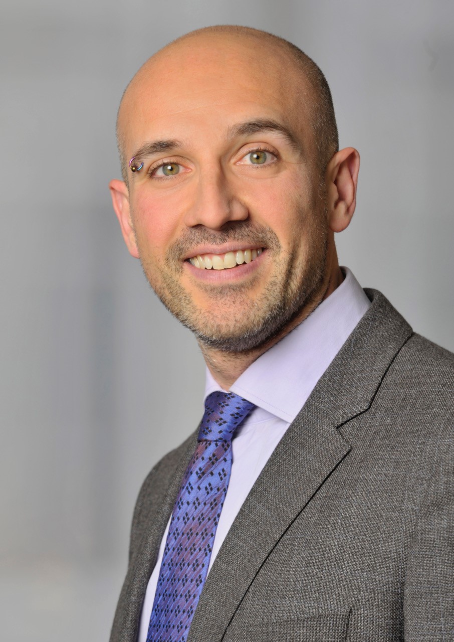
Dr. Alessio Attardo
alessio_attardo@psych.mpg.de
https://www.psych.mpg.de/2037836/attardo
https://sites.google.com/view/the-a-lab/home
The capability to store and consciously recall past experiences is crucial for survival and establishing a coherent sense of reality. This ability in mammals is dependent on the hippocampal formation, which encodes experience through coordinated neuronal activity enabling memory acquisition and recall. Our group is interested in the cellular mechanisms underlying the hippocampal ability to encode experience and memory. Furthermore, we are interested in how stress response impairs such an ability leading to temporary and permanent memory impairment. We use two-photon optical imaging as well as wide field head mounted miniaturized microscopes to study neuronal structural plasticity and experience representation in the hippocampal CA1 of live mice performing hippocampal-dependent tasks. We also take advantage of molecular and genetic tools to investigate defined synapses and neuronal subpopulations and manipulate activity of defined neuronal populations. Together these tools enable us to investigate the interplay between cellular events and learning in the hippocampal formation going from molecular mechanisms to network-level computation.
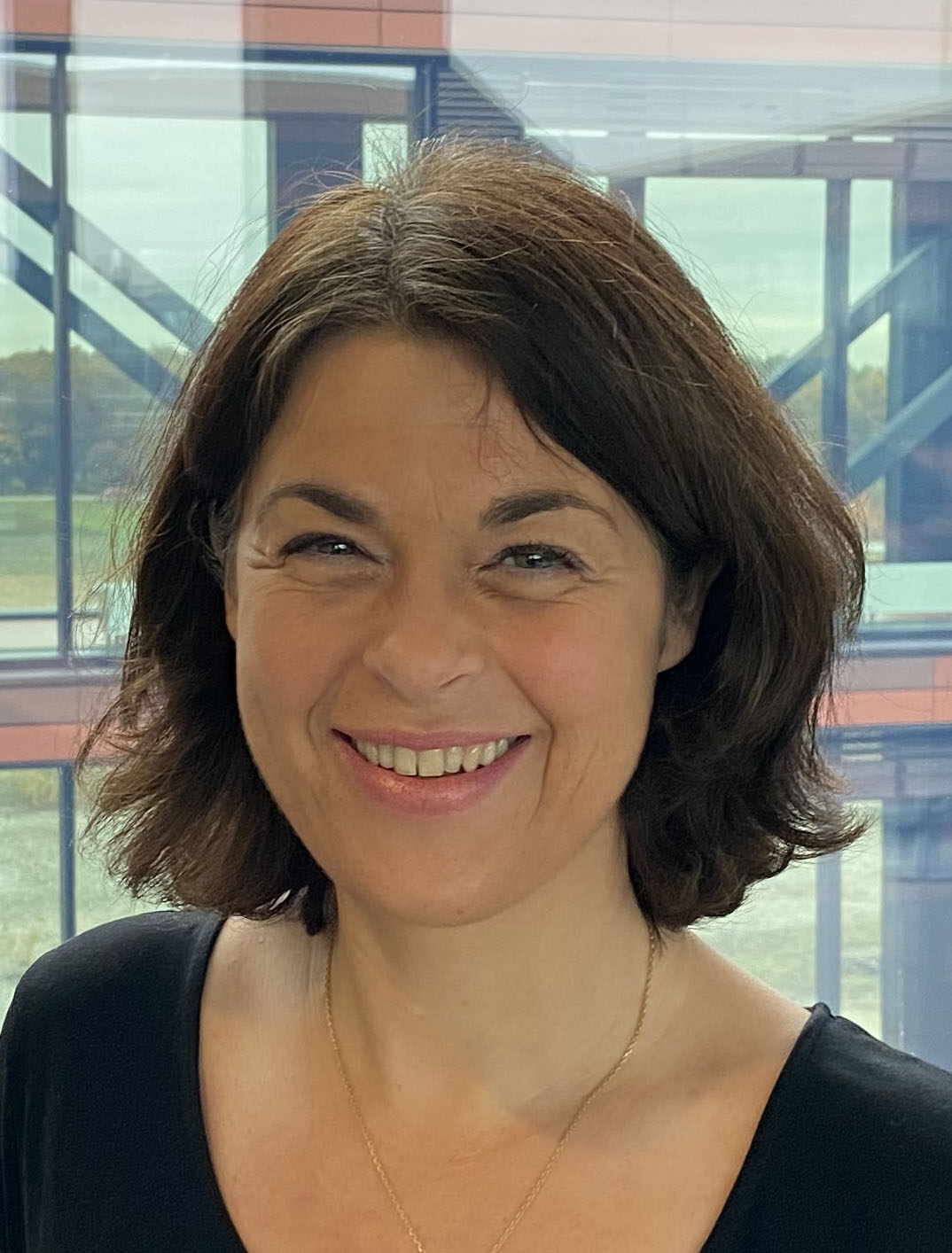
Dr. Florence Bareyre
Florence.bareyre@med.uni-muenchen.de
Neuroimmunology Munich | Bareyre Lab
Traumatic, ischemic and inflammatory lesions to the spinal cord lead to the transection of descending and ascending axonal tract systems. If these lesions are complete – i.e. if all axons in the spinal cord are transected – severe and persistent functional deficits ensue. If however the lesions are incomplete and some axonal tracts are spared, some recovery of function can be observed. We are studying the anatomical, functional and molecular mechanisms underlying the recovery process in an attempt to develop new therapeutic strategies that can support spinal cord repair in neurological disease caused by trauma, ischemia or inflammation. Our lab is currently investigating the regulation of synapse formation and elimination following spinal cord injury, the activity-dependent regulation of axonal plasticity following spinal cord injury as well as the neuronal regulation of inflammation following spinal cord injury. In addition to spinal cord injury, we are also interested in the acute and long term effects of mild repetitive traumatic brain injury.
Michael Josef Brunnhuber
michael.brunnhuber@tum.de
http://www.mec.med.tum.de/en/michael-brunnhuber
Our group develops and implements innovative teaching and learning strategies based on education research. Our mission is to improve and advance pedagogy and education at university level. We are active in the field of education and education research in natural sciences, mathematics and medicine.
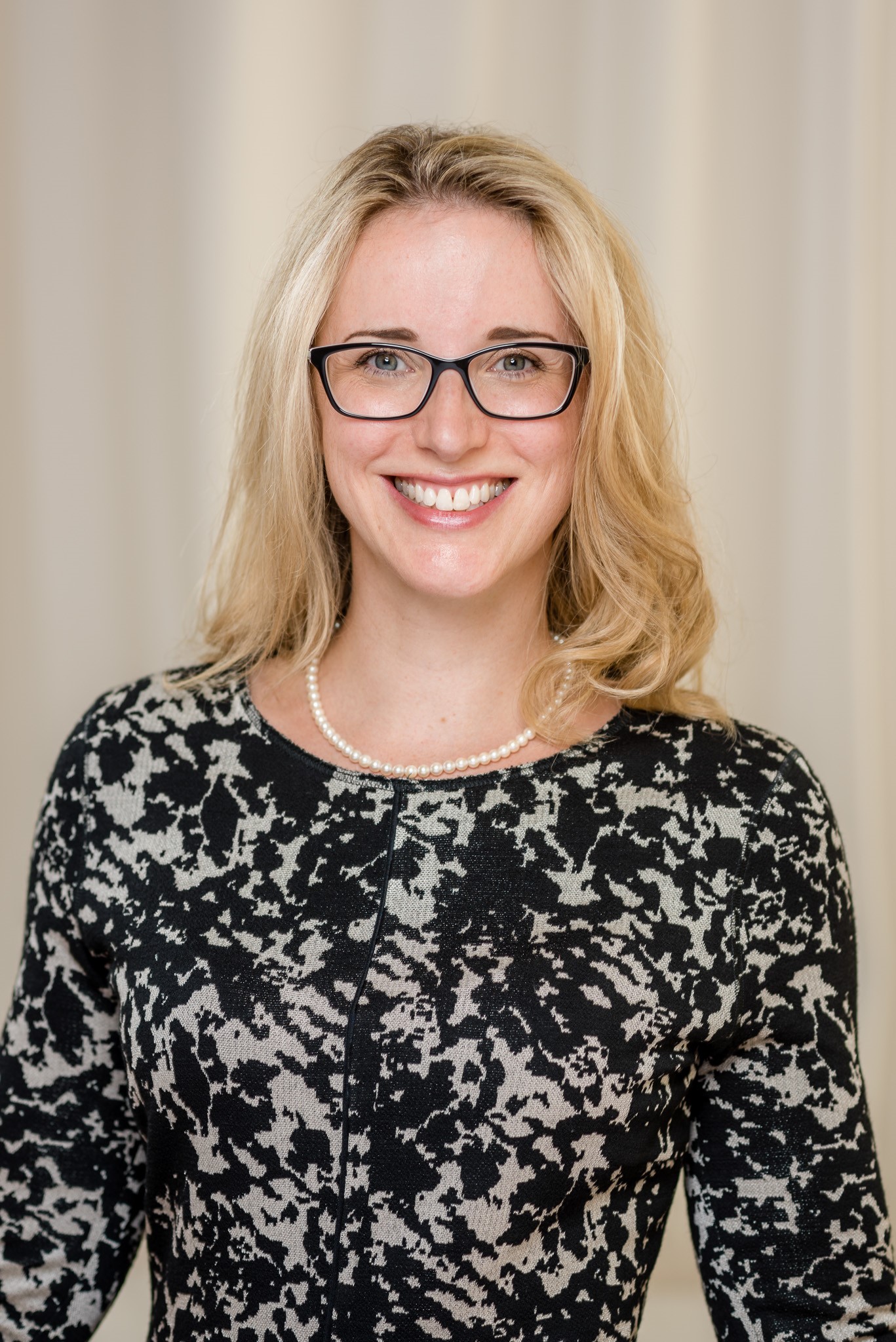
Prof. Dr. Alena Buyx
medizinethik.med@tum.de
http://www.get.med.tum.de/
https://www.professoren.tum.de/en/buyx-alena/
The Institute for the History and Ethics of Medicine works on the ethical, historical and cultural dimensions of medicine. In medical ethics, we focus on questions of responsible development and ethical integration of new biomedical technologies into research and clinical practice. Current examples are data-driven technologies such as biomarkers, applications in embodied AI/robotics, or neurostimulation in children. Further focal points are new concepts of solidarity in medicine as well as issues in neuroethics, research ethics and public health ethics. The ethics group at the Institute takes an interdisciplinary approach. We conduct various mixed-methods studies (qualitative, quantitative and theoretical methods) and we strive for an embedded approach to developing and ‘co-creating’ novel biomedical innovations, where clinicians, engineers, programmers etc. and ethicists and social scientists work side by side to embed ethical and social considerations into the development process. We also take an active interest in public engagement about ethical and social issues in biomedicine, through public events, consulting for large research consortia, or by advising policy-makers (WHO, German Ethics Council etc.).
Prof. Dr. Cristina García Cáceres
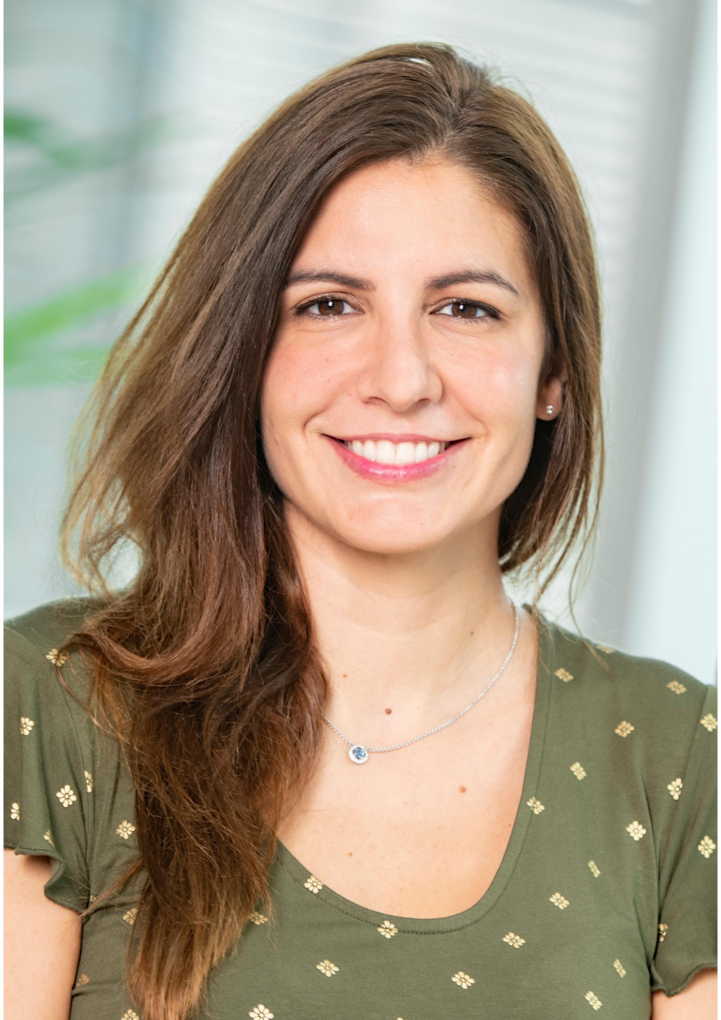
garcia-caceres@helmholtz-muenchen.de
Cristina García-Cáceres is a W2 professor at the Faculty of Medicine at the LMU and was originally recruited to the Helmholtz Munich in 2012 from the Universidad Autónoma de Madrid as a Postdoctoral Scientist. In 2015 was promoted to lead the Astrocyte-Neuron Network Unit at the Institute for Diabetes and Obesity and since then she established and consolidated her own research group performing cutting-edge, state-of-the-art experiments in collaboration with internationally recognized leaders in the field of neuronal, glial cell biology and obesity. Her work was awarded by the prestigious ERC Starting Grant and has already resulted in breakthrough discoveries highlighting that hypothalamic circuits controlling feeding and metabolism require a precise, finely-tuned and coordinated, communication between astrocytes and neurons for maintaining a balanced systemic metabolism and blood pressure. Indeed, her findings represents a paradigm shift in how glucose gets into the brain (Cell 2016) as well as highlight unprecedented role of astrocytes in tuning sympathetic outflow towards cardiovascular target organs (Cell Metab 2021).
Prof. Dr. Janine Diehl-Schmid
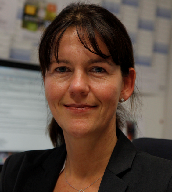
janine.diehl-schmid@tum.de
https://www.ncbi.nlm.nih.gov/sites/myncbi/1bMwrpKSXyHAJ/bibliography/483...
Janine Diehl-Schmid, MD, is a board certified psychiatrist and psychotherapist. She is employed at the Department of Psychiatry and Psychotherapy of Technical University of Munich, where she serves as a senior consultant and is co-heading the Center for Cognitive Disorders. Her particular research interests include frontotemporal dementia, young onset dementia and, more recently, palliative care in dementia. She has authored and co-authored more than 140 peer-reviewed articles with a cumulative Impact Factor of app. 500.
PD Dr. Thomas Fenzl
Thomas.fenzl@tum.de
www.anaesth.med.tum.de
www.gsn.uni-muenchen.de/people/faculty/associate/fenzl
We are interested in interactions between sleep and anesthesia within the mammalian brain. Does sleep influence particular states of anesthesia, does anesthesia interfere with subsequent sleep, does anesthesia impair memory consolidation during sleep, does sleep decrease potential cognitive deficits after anesthesia and, as a very fundamental question, what happens within the brain at a systemic level when this organ switches between sleep, anesthesia and wakefulness? Our newly established lab applies highly sophisticated methods in computer programming, behavioral biology and in vivo electrophysiology and combines the knowledge of biologists/neuroscientists, computer experts and clinicians under one roof.
PD Dr. phil. habil. Martin Gartmeier
martin.gartmeier@tum.de
http://www.mec.med.tum.de/de/pd-dr-phil-habil-martin-gartmeier
Martin Gartmeier, PhD, (born 1976) is Research Coordinator at the Chair of Medical Education (Prof. Dr. Pascal Berberat) at the TUM School of Medicine, Klinikum Rechts der Isar. His research interests are professional learning and professional competence, especially in the area of communication in clinical contexts. Martin Gartmeier earned his PhD at the University of Regensburg through researching how employees from different vocational domains learn from errors and how such learning affects their professional knowledge. Later, he worked at the TUM School of Education in an interdisciplinary project which focused upon fostering professional communication competence in the domains of teacher and medical education.
Dr. Leanne Godinho
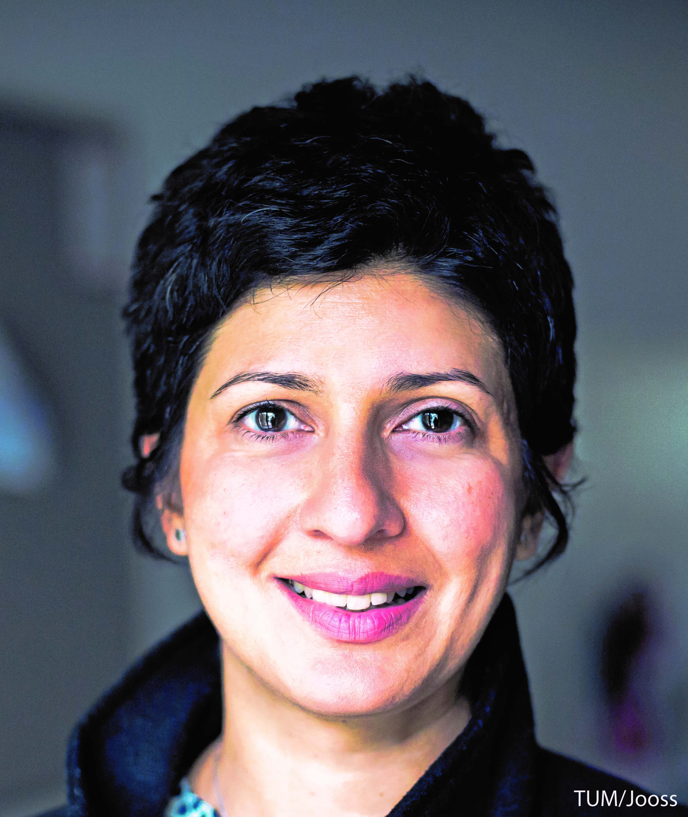
leanne.godinho@tum.de
https://web.med.tum.de/neuroscience/godinho-lab/
We are interested in understanding how the vertebrate central nervous system (CNS) is assembled during development and exploit the retina, an accessible part of the CNS to do so. We use zebrafish, highly visual vertebrates that develop ex utero and are largely translucent during development, to study retinal development in real time in vivo. We probe the mechanisms that underlie the generation of diverse cell-types, their differentiation and integration into synaptic circuits. By combining genetic tools and in vivo time-lapse imaging, we can follow the developmental trajectories of cells from the time of their ‘birth’ to their arrival at their definitive locations, and their integration into the local circuitry.
Prof. Dr. Ilona Grunwald Kadow
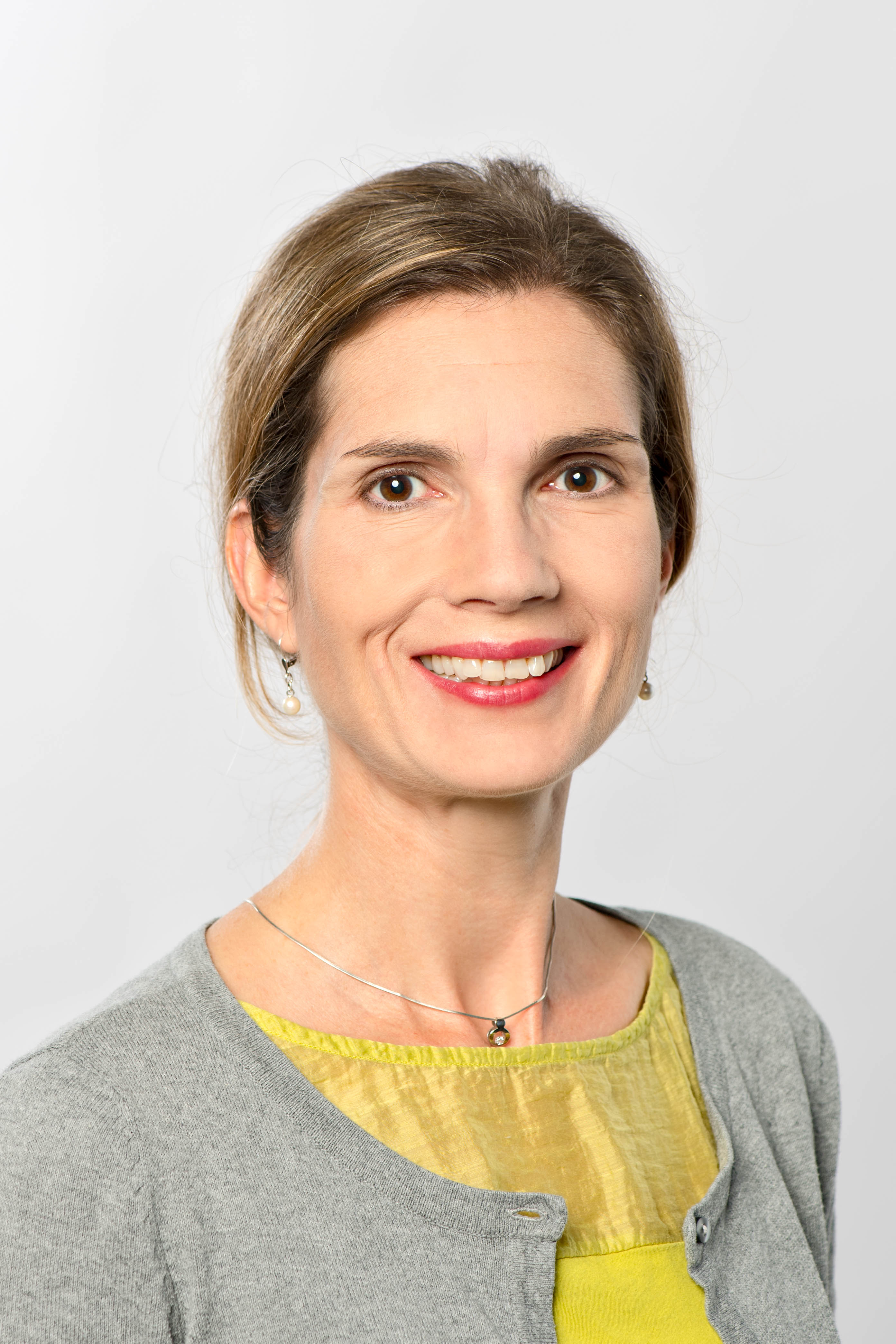
ilona.grunwald@tum.de
http://neuro.wzw.tum.de/index.php?id=1
We aim at answering these questions at three levels: (1) behavior, (2) neural networks, and (3) genes. To this end, we are using genetic models such as Drosophila melanogaster in combination with modern techniques including high resolution behavioral analysis, optogenetics, and in vivo multiphoton microscopy. In particular, we focus on how the brain translates chemosensory information, i.e. odors and tastes, into state- and experience-dependent perceptions and ultimately into behavior.
Prof. Dr. Simon Jacob
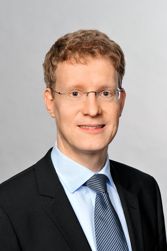
simon.jacob@tum.de
www.simonjacob.de
The Jacob laboratory at the Department of Neurosurgery studies the cellular mechanisms underlying higher cognitive functions. We are particularly interested in how various brain regions interact to enable intelligent, goal-directed behavior. Our current focus is the role of subcortical neuromodulators such as dopamine in regulating cortical circuits that are involved in perception and decision-making. Disrupted neuromodulator signaling is heavily involved in the generation of neuropsychiatric disorders, and we work towards a better understanding of the cellular basis of many mental diseases. We use a variety of techniques in awake trained mice, including behavioral analyses, large-scale extracellular recordings, optogenetic manipulation of defined cell types and networks, fluorescent imaging and computational modeling. In a special translational approach, we are also recording from individual neurons in human neurosurgical patients.
Prof. Dr. med. Jan Kirschke
jan.kirschke@tum.de
jan.kirschke@mri.tum.de
https://www.neurokopfzentrum.med.tum.de/neuroradiologie/347.html
Jan Kirschke is attending radiologist at the department of neuroradiology at the Technische Universität München (TUM) in Munich, Germany. He is working on quantitative imaging biomarkers for spine and neuro applications. Within his ERC-funded spine research group, he works on multimodal imaging approaches combined with biomechanical modeling and machine learning to predict fragility fractures and back pain. This includes data science research to handle huge datasets and create structured medical data. He also investigates imaging biomarkers to assess local brain tissue properties in brain tumors and MS for objective and accurate disease assessment and prediction.
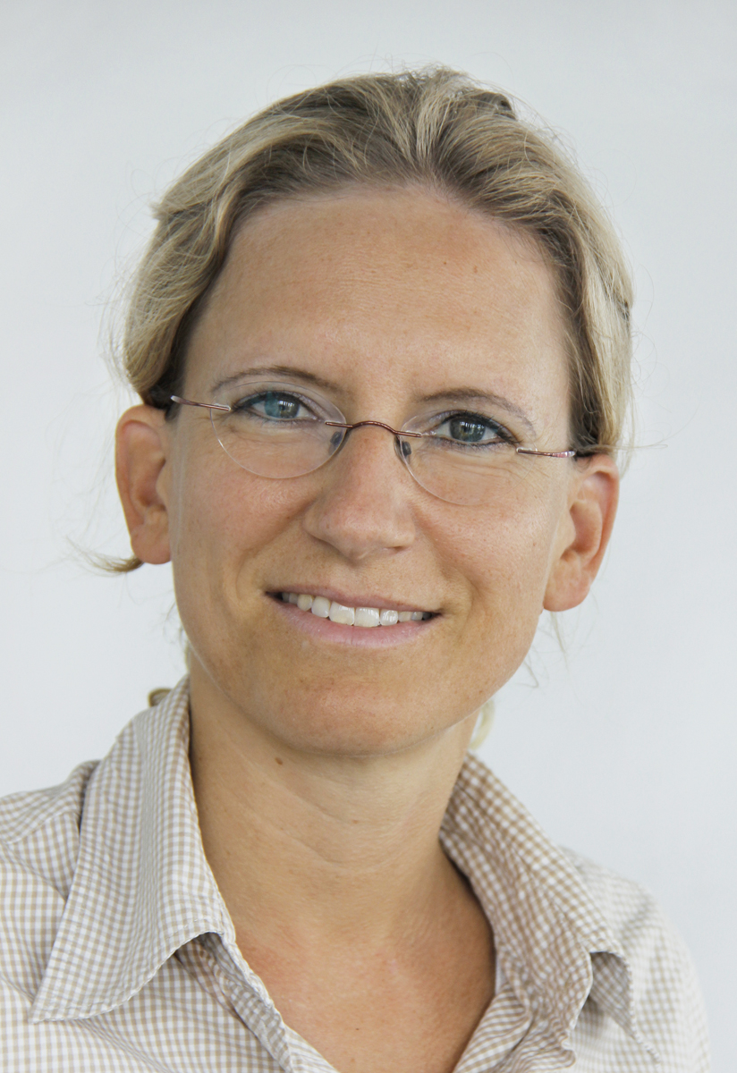
Prof. Dr. Kathrin Koch
kathrin.koch@tum.de
https://www.neurokopfzentrum.med.tum.de/neuroradiologie/forschung_projek...
We investigate normal and altered brain function in healthy humans and neuropsychiatric patients. In this context we are mainly interested in the effects of different interventions (e.g., cognitive training, mindfulness training, tDCS) on functional activation as well as functional and structural connectivity in healthy people and in finding out more about functional and structural alterations going along with psychiatric disorders such as obsessive-compulsive disorder (OCD). To this aim, we combine different imaging methods including DTI, functional MRI and PET to study white matter brain structure, brain function in the resting state and during task as well as brain metabolism and their changes following specific interventions or in association with psychiatric symptoms and conditions.
Prof. Dr. Thomas Korn
thomas.korn@tum.de
https://kornlab.med.tum.de
Fate decisions of T cell in vivo: Our major focus is the functional phenotype of T cells in vivo. T cells are key organizers of immune responses to foreign and self antigens. They determine the specificity of an immune response but also the quality and strength of the inflammation that accompanies an immune response. T cells are very motile cells characterized by long range trafficking across multiple compartments. We want to understand the cues that trigger differentiation processes and effector functions of T cells and the downstream molecular pathways. Our particular interest is to analyze to whom and in what language T cells talk in distinct non-lymphoid microenvironments. Our main preclinical model systems focus on autoimmune inflammatory desease paradigms (Multiple Sclerosis and Neuromyelitis optica), but our approaches are also applicable to chronic inflammation and tumor immunity.
Dr. rer. nat. Matthias Kreuzer
Prof. Dr. Sandro Krieg
sandro.krieg@tum.de
sandro.krieg@mri.tum.de
http://www.tumnic.mri.tum.de/tumnic/x1.html
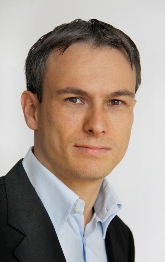
Sandro Krieg is an attending neurosurgeon but also group leader of the Department of Neurosurgery at the Technichal University of Munich. He specializes in the diagnosis and treatment of eloquent brain tumors including pre- and intraoperative mapping techniques. After a stay at UCSF San Francisco he further refined his research group, which is mainly focussing on the development of new protocols and techniques of pre- and intraoperative mapping of neurological function. This include navigated transcranial magnetic stimulation (nTMS) and direct electrical stimulation of the cortical and subcortical brain and the application of refined imaging techniques such as nTMS-based tractography and connectomic analyses.
Dr. Monika Leischner-Brill
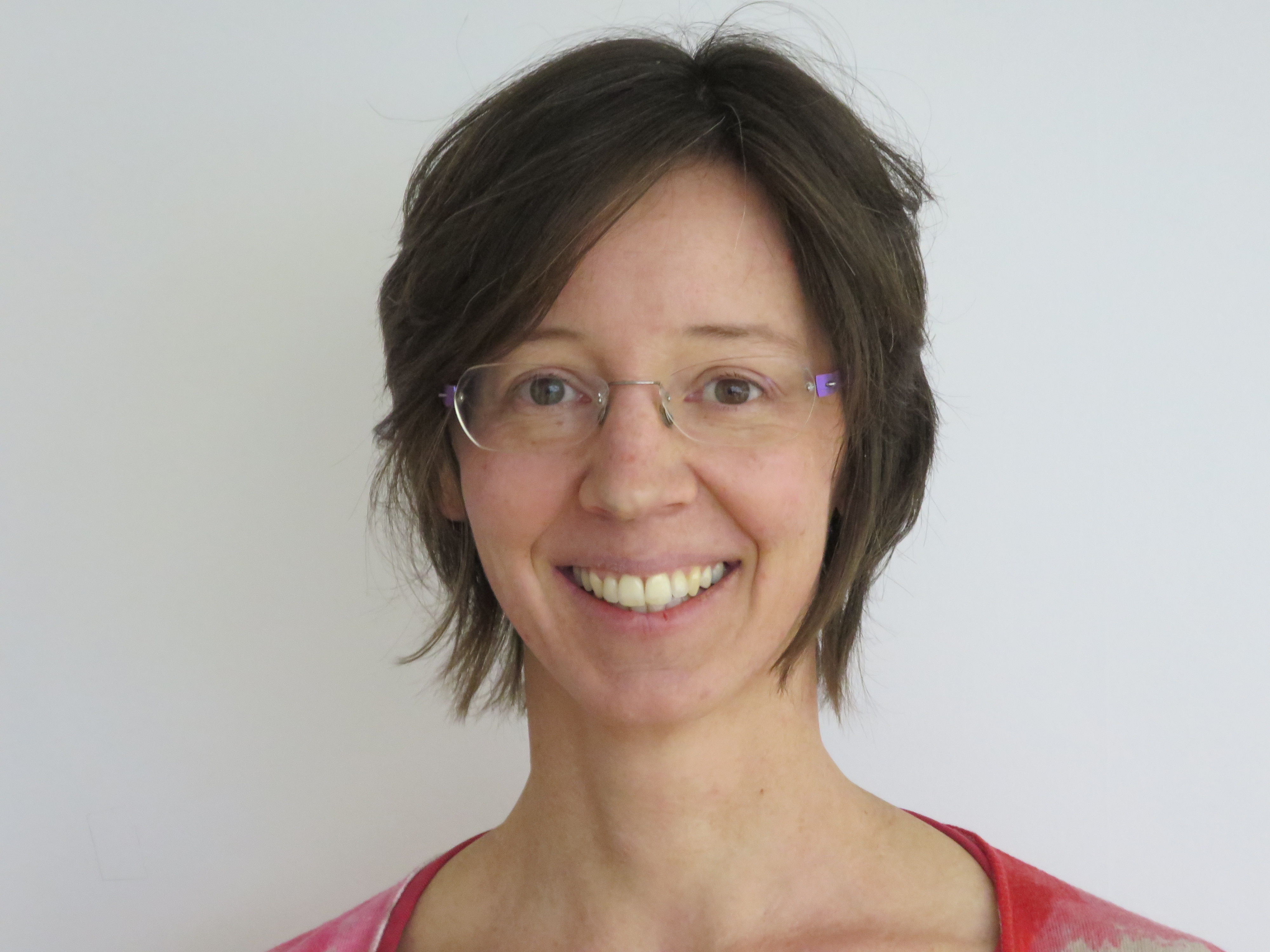
monika.leischner-brill@tum.de
https://www.neuroscience.med.tum.de/index.php?id=24&L=0
We are interested in the cell biology of synapse elimination, a developmental process where exuberant connections are removed to ultimately form a precisely wired neural circuit. As a model, we look at murine motor axons and sensory neurons, combining genetic tools and time-lapse imaging in nerve-muscle explants. When axons are pruned back, the cytoskeleton needs to be dismantled, while it stabilizes in branches, which are maintained life-long. We study changes of the cytoskeleton during the synapse elimination process, especially microtubule, as organelles and vesicles are transported along them with the help of molecular motors. We focus on microtubule dynamics and their post-translational modification profile, which can be added and removed enzymatically and changes in pruning axons. One post-translational modification attracts spastin, a microtubule severing enzyme and genetic ablation of spastin leads to microtubular stabilization. Spastin mutation in humans lead to the neurodegenerative disease hereditary spastic paraplegia (HSP), where neurons with long axons are prone to degeneration. To transfer our findings from mouse motor axons to humans, we recently started to look at human skin biopsies from HSP-patients.
Prof. Dr. Stefan Lichtenthaler
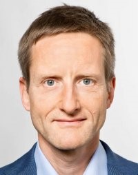
stefan.lichtenthaler@dzne.de
www.dzne.de/lichtenthaler
We study how Alzheimer’s disease develops in the brain on the molecular and cellular level. The aim of our research is to better understand the disease causes and to develop new diagnostic, therapeutic and preventive approaches. Additionally, we want to predict possible side effects of Alzheimer-targeted drugs and, thus, make drug development safer. For our interdisciplinary research we use a variety of modern methods from biochemistry, proteomics, molecular, cellular and neurobiology as well as in vitro and in vivo models of Alzheimer’s disease. The focus of our research is on the molecular scissors (proteases) ADAM10 (alpha-secretase), BACE1 (beta-secretase) und gamma-secretase as well as microglia-dependent inflammatory processes in the brain. These molecules and processes have a central role in Alzheimer’s pathogenesis. We investigate their physiological function in the healthy brains and develop ways to specifically target them for a treatment of Alzheimer’s disease. Additionally, we develop proteomic methods for a faster and more detailed study of these proteases, but also of the brain’s cerebrospinal fluid. This will not only allow a better understanding of the brain, but also help to develop new diagnostics for evaluating whether patients respond well to a drug.
Prof. Dr. Arthur Liesz
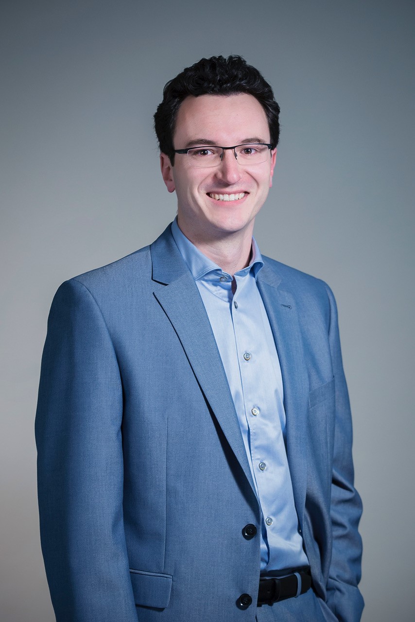
Arthur.Liesz@med.uni-muenchen.de
https://www.isd-research.de/liesz-lab
We are interested in the interplay between the brain and the immune system after stroke. Acute brain lesions disturb the well-balanced interconnection between both systems. Hence, our research focuses on both directions of brain-immune interaction: The impact of immune mechanisms on neuronal damage and recovery and the systemic immunomodulation after stroke. Our methodological spectrum covers diverse brain ischemia models, transgenic animal models, a broad spectrum of cutting-edge immunological techniques as well as histological, biomolecular and behavioral analysis tools. The lab has a strong translational research focus with the ultimate goal to develop novel diagnostic tools, therapies and mechanistic insights on the highly complex disease which stroke represents. In order to achieve this, a premise of our work is to address key unmet needs of stroke patients and make use of translationally relevant tools.
Prof. Dr. Paul Lingor
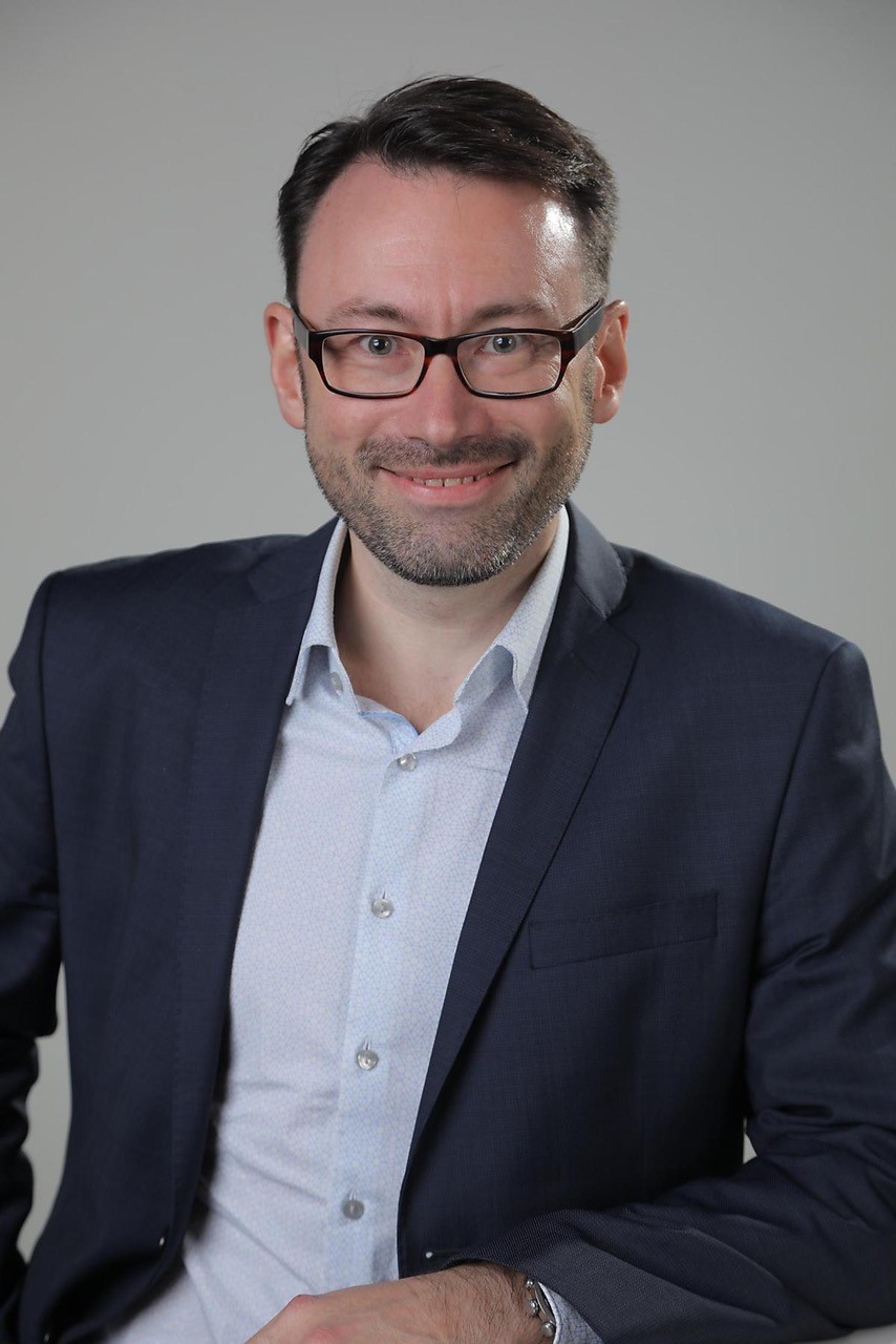
paul.lingor@tum.de
http://www.neurokopfzentrum.med.tum.de/neurologie/42lingor.html
Being a clinical neurologist, the motivation of my work is derived from clinical problems in the diagnosis and treatment of neurodegenerative disorders, particularly Parkinson’s disease (PD) and Amyotrophic lateral sclerosis (ALS). My group uses basic neurobiological techniques to understand the mechanisms of the underlying pathophysiology in degenerative CNS disorders, i.e. synapto-axonal degeneration or iron-mediated neurotoxicity. Body fluids, such as CSF, blood or tear fluid are collected and used by our group to identify novel biomarkers for better diagnosis and subgroup stratification. In a translational approach, we develop novel therapeutic strategies for neurodegenerative disorders bridging the gap from animal studies to human clinical trials.
Prof. Dr. Dominik Paquet
dominik.paquet@med.uni-muenchen.de
https://www.isd-research.de/paquetlab
We are interested in the molecular and cellular mechanisms leading to neuronal death and cognitive decline in patients with neuropsychiatric disorders (e.g. Alzheimer’s disease and Frontotemporal dementia) and neurovascular impairments (stroke and vascular cognitive impairment). Our main focus is to use human induced pluripotent stem cells (iPSCs) to build brain tissue models recapitulating these diseases. We have recently developed efficient technologies to introduce and remove patient mutations in iPSCs using the CRISPR/Cas9 gene editing system and have also developed protocols for the optimized differentiation of all major cell types of the human brain. We apply these technologies to generate human cortical neurons and other brain cells with mutations in disease-associated genes (such as APP, PSEN or TAU) and isogenic controls, and then combine these cells into human brain tissue models to elicit disease-relevant phenotypes and reveal molecular disease mechanisms.
Prof. Dr. Markus Ploner
markus.ploner@tum.de
https://www.painlabmunich.de/
We investigate how the human brain subserves the experience of pain. To this end, we use electroencephalography to record brain activity in healthy human participants and patients suffering from chronic pain. As we are particularly interested in the functional significance of neuronal oscillations and neuronal communication we perform complex time-frequency and connectivity analyses of brain activity. Moreover, we use non-invasive brain stimulation and neurofeedback to modulate neuronal oscillations. The ultimate goal of our work is to further the understanding, diagnosis and therapy of chronic pain.
Prof. Dr. Ruben Portugues
rportugues@neuro.mpg.de
https://www.neuro.mpg.de/portugues
In our lab we study how the brain processes sensory stimuli and uses these to select and improve its behavior. We use a simple vertebrate model organism, the larval zebrafish, because it gives us the opportunity of studying this question at multiple scales: from single neurons to the whole brain. Zebrafish are vertebrates, and as such, share a similar brain plan to that of mammals. We use a variety of techniques that range from exhaustive quantitative behavioral analysis and psychophysics to monitoring physiological neuronal activity using genetically encoded calcium indicators and electrophysiological whole-cell recordings and include optogenetics, theoretical and computational models and machine learning and pharmaco-genetic tools.
PD Dr. Christine Preibisch

preibisch@tum.de
https://www.neurokopfzentrum.med.tum.de/neuroradiologie/3216.html
The Magnetic Resonance (MR) Physics group at the Department of Neuroradiology at the Technische Universität München (TUM) is interested in exploiting a variety of different MR contrast mechanisms to obtain functionally relevant physiological parameters via magnetic resonance imaging (MRI). Core areas of research comprise quantification of blood oxygenation and perfusion and application of those techniques in multimodal MRI studies. Currently, we are expanding our methodological portfolio towards quantitative structural MRI and simulation techniques. The overarching goal of our work is to elucidate physiological mechanisms of brain function in health and disease and their interplay with structural and functional impairments.
PD Dr. Valentin Riedl
valentin.riedl@tum.de
https://valentinriedl.de
https://www.professoren.tum.de/en/tum-junior-fellows/r/riedl-valentin/
Neuroenergetics research group: Neuronal communication among highly connected brain regions is the main driver of the brain’s energy demands. While we know much about the macroscopic organization of the human brain in specialized regions and brain networks, the energy budget of brain function is still unclear. Moreover, brain metabolism is heavily disturbed in several neuropsychiatric disorders but the relationship to brain network communication is also unknown. In my research group, we measure the brain's energy consumption and relate these to common measures of brain organization. Methods: We simultaneously acquire energy metabolism and brain connectivity measures on an integrated PET/MR scanner. We measure inhibitory and excitatory neurotransmitter levels using 1H-Magnetic Resonance Spectroscopy (MRS) and modulate brain function using non-invasive, stereotactic transcranial magnetic stimulation (TMS). Research projects: - Develop new approaches to integrate brain profiles of energy metabolism and network connectivity. - Study the energy metabolism of brain networks during memory consolidation and modulate this process with non-invasive brain stimulation. - Link brain energetics with nutrition and body metabolism. - Uncover deficient metabolic brain profiles in patients with neuropsychiatric disorders.
Dr. Martina Schifferer
martina.schifferer@dzne.com
https://www.synergy-munich.de/members/associate-investigators/schifferer/index.html
The Nanoscale hub collaborates with different neurobiology research groups by applying Electron Microscopy (EM) and focuses on technical developments. Ultrastructural analysis not only provides unprecedented resolution but reveals reveal additional, non-hypothesis-driven insights. Typical examples comprise the characterization of neuronal, organellar, myelin, fibril or blood vessel morphologies or their context within tissue, including pathological states like MS, AD and stroke. Currently, model organisms include mouse, zebrafish and human biopsies. The hub provides room temperature Transmission (TEM) and Scanning EM (SEM) covering all steps from sample processing, image acquisition to image analysis. Advanced methods like Correlative Light and Electron Microscopy (CLEM) and Volume EM (FIB-SEM, ATUM) are tailor-made and further developed.
Dr. Barbara Schormair
barbara.schormair@tum.de
https://www.helmholtz-muenchen.de/en/ing/index.html
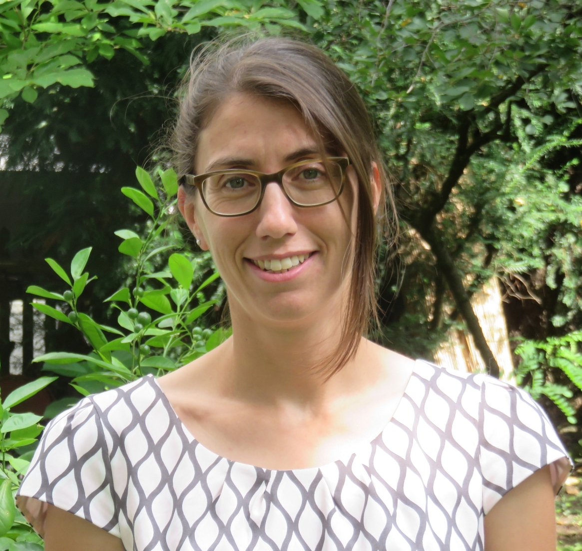
Barbara Schormair studied molecular biology and focused on human genetics during her PhD thesis. She is deputy director of the Institute of Neurogenomics (ING) at the Helmholtz Zentrum München. Her main research interest is the application of genomic technologies to understand the genetic basis of Restless Legs Syndrome, one of the most common neurological disorders. The ING is dedicated towards deciphering the underlying genetic basis of movement disorders and turning this knowledge into improved patient care by combining classical wet lab work, high-throughput omics technologies, and computational approaches.
Moritz Schumm
moritz.schumm@tum.de
https://www.mec.med.tum.de/de/moritz-schumm
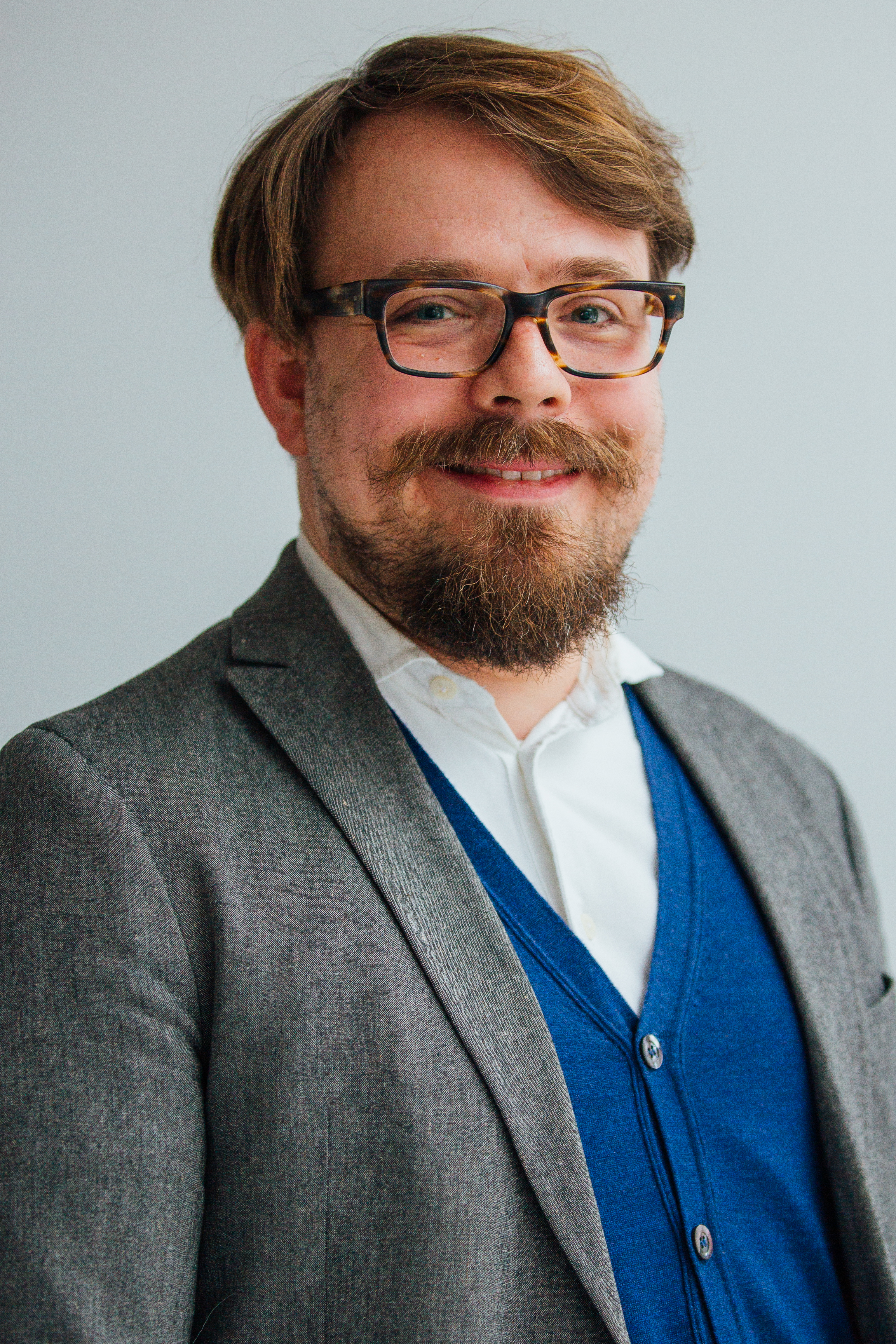
The Medical Humanities Education at TUM Medical School aims at stimulating, encouraging and supporting medical students to develop and cultivate a personal professional identity that is based on a far-reaching critical understanding of what medicine and life sciences can and/or have to be concerning the different dimensions of the human subject. To achieve this we seek to incorporate humanities, literature, film and arts into our curriculum and develop custom-made lectures, courses and workshops.
PD Dr. Christian Sorg
christian.sorg@tum.de
http://www.tumnic.mri.tum.de/tumnic/x1.html
The Neuropsychiatry and Neuroimaging Lab studies large-scale brain systems in healthy humans and neuropsychiatric patients by the use of multi-modal imaging techniques. Main focus is on neurodevelopmental disorders such as schizophrenia and premature birth. Key method is functional MRI extended by EEG, perfusion-based MRI, and PET. We are interested in how blood oxygenation fluctuates – i.e. the main functional MRI signal -, how these fluctuations link with both neural oscillations and blood perfusion, how they spread across the cortex, and how these processes are impaired in developmental disorders. Therefore, we use simultaneous in-vivo imaging of ongoing brain processes at rest in humans together with a modelling approach on processes of interest.
Prof. Dr. Gil Westmeyer
gil.westmeyer@tum.de
https://www.professoren.tum.de/en/westmeyer-gil/
https://www.westmeyerlab.org/
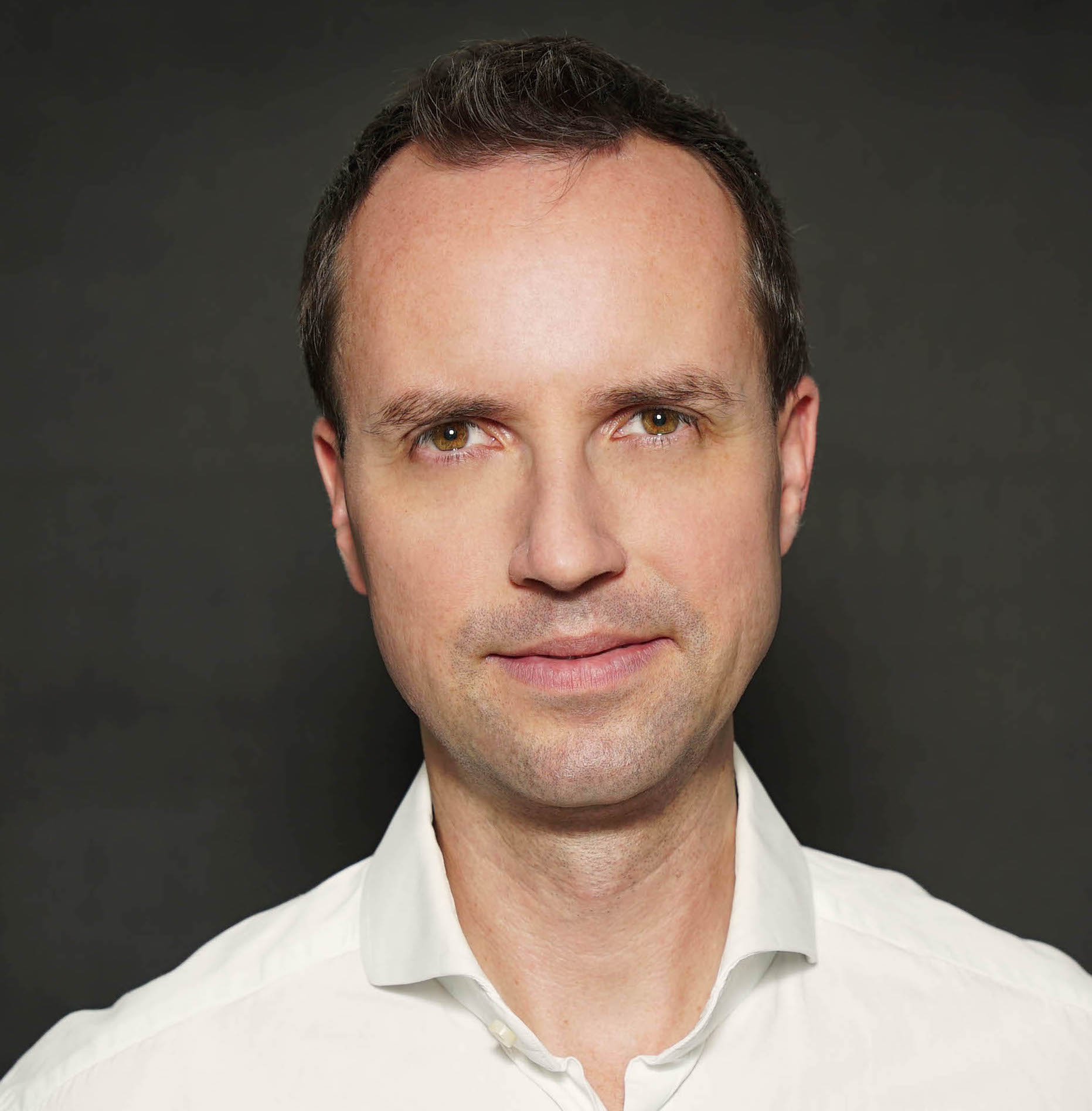
We develop new molecular interfaces to interrogate neuronal processes across scales: we, eg., work on genetically encoded reporters for Electron Microscopy (EM), we build techniques for longitudinal monitoring of local isoform expression, and we generate biomagnetic interfaces that can be read-out via Magnetic Resonance Imaging (MRI) and actuated via magnetic fields. We also develop molecular sensors for combined fluorescence and optoacoustic neuroimaging in (freely-swimming) zebrafish and rodent models to decipher long-range neuronal activity in response to controlled perturbations. As a long term goal, we seek to combine these molecular sensors and actuators to enable closed-loop control of simple neuronal circuits.
PD Dr. med. Benedikt Wiestler
b.wiestler@tum.de
https://compimg.github.io
Advances in AI have fostered the development of machine learning models able to master medical image analysis tasks that were previously thought unsolvable for computers. Within our working group "Computational Imaging" we develop such models in close interdisciplinary collaboration between medicine and computer science. Focusing on two model brain diseases (Multiple Sclerosis and Gliomas), our research spans the entire imaging work flow, from image synthesis to segmentation and modelling of disease. Ultimately, we are aiming to make the wealth of information about a patient's disease, which is hidden in medical imaging data, available for improved patient care.

